
Mother of Nudibranchs: Raising the Hooded Nudibranch
I remember sitting in a lecture hall on one Fall day in 2013 when I was asked, “What’s your favourite invertebrate?”. This wasn’t such a weird question, as the class I was in was Biology 321: Survey of Invertebrates at the University of Victoria (UVic) in BC, Canada. My friend sitting beside me shared that hers were gastropods, or more specifically, nudibranchs. My first reaction was shock with a slight touch of horror. Why snails?! At that point in time, I was strictly interested in sea urchins, as it was the purple sea urchin that I held in high school that made me realize I wanted to study Marine Biology. Coincidentally, it was on that day in BIOL 321 that we launched into a lecture on gastropods, and I was introduced to the wonderful world of nudibranchs.
Now, if you don’t know what a nudibranch is, it is imperative that you google it right now. What you will find is a world of diversity in shape, size and colour. Some of these animals literally look like Pokémon. Nudibranchs are marine snails that live without a shell. That’s right, in an ocean full of animals trying to eat you these bad B’s are living without the one protective retreat snails typically have. Instead, nudibranchs have multiple alternate defensive techniques such as swimming or absorbing the toxic chemicals or structures from their food sources to make themselves taste bad or downright dangerous to predators. Another defensive technique is the ability to autotomize or “self-cut” an appendage. Think of a lizard’s tail, or a crab’s pincher breaking off; the ability to autotomize is surprisingly prevalent in the animal kingdom. This was the defensive technique that I studied in the Summer of 2014 in the species Melibe leonina, the Hooded Nudibranch.

Adult Hooded Nudibranch. Picture courtesy of Robin Agarwal - Own work, CC BY-SA 4.0
This wonderful work opportunity was with Dr. Louise Page, the same instructor from BIOL 321. It turned out that Dr. Page was a snail lover too, with a particular interest in the development and evolution of marine snail species, including nudibranchs. My goal was to raise the young of the Hooded Nudibranch and figure out at what point in development they were capable of autotomizing their appendages.
The Hooded Nudibranch, is an inhabitant of eelgrass beds in the intertidal zone along the West Coast of North America, ranging from Alaska to California. This species is particularly obvious with its very large oral hood (hood with tentacles resembling a lion’s mane) that is used for capturing prey, translucent body and paddle-like appendages called cerata (singular: ceras). Crabs are one of the main predators of the Hooded Nudibranch, so the ability to autotomize a ceras if a crab grabs on is an excellent escape technique. In fact, these nudibranchs typically autotomize as a distraction and then swim away! The perfect one-two punch to get the heck out of there. The real question is: when do these animals become capable of autotomy during their development?
To determine this we collected the white, ribbon-like egg masses of the Hooded Nudibranch at Patricia Bay, Vancouver Island, BC, and brought them back to the Page Lab at UVic. There, we submerged the egg masses in locally collected sea water and waited for the larvae to hatch out. That’s right, the first stage in a marine snail’s life cycle is typically a larva called a veliger. Veliger larvae are microscopic, beautiful little creatures that swim around eating algae until they are big enough to settle on the seafloor. Once they encounter their optimal habitat, they undergo metamorphosis to become a juvenile, much like a caterpillar turning into a butterfly. For this species, the larval stage lasted around 30 days. So, for one month I painstakingly cared for these little larvae, feeding them algae and giving them fresh water. This was a lot more complicated than it sounds since I had to grow the algae (which required attention every week), collect seawater, filter and treat the seawater in case of excess heavy metals, and pipette each of the hundreds of larvae into fresh water bowls every couple of days. These larvae felt quite literally like my children.

Larvae of the Hooded Nudibranch. A. Hundreds of small larvae, appearing as small beige specks, in a 500 mL beaker. B. Magnified larva showing its swimming apparatus (top with hair-like cilia) and round shell. Because they are quite transparent you can also see some of the internal organs. Pictures courtesy of Nova Hanson and the Page Lab.
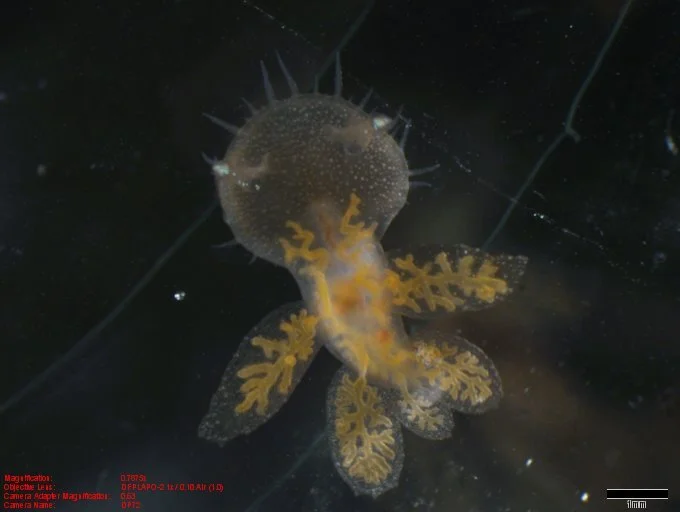
Late Stage 3 Juvenile of the Hooded Nudibranch showing developed oral hood (top) and numerous ceratal appendages which are capable of autotomy. Scale bar 1 mm. Picture courtesy of Nova Hanson and the Page Lab.
Once they were about a month old, the veliger larvae settled on the bottom of the bowls and underwent metamorphosis, changing drastically in appearance to become juveniles that looked like miniature adults. Previously, Dr. Page characterized the developmental stages of the Hooded Nudibranch, publishing work that defined the five stages of juvenile development (Bickell-Page 1989), mainly based on the size of the juvenile and number of cerata. After metamorphosis, one by one the nudibranchs grew bigger and their cerata began to develop. With the change from veliger larva to juvenile also came a change in diet. These juveniles were carnivores that fed by trapping small crustaceans under their oral hood and sucking them into their mouth. This led me to collecting larval crustaceans so that I could feed them to my nudibranch children. As they grew in size and number of cerata over the next couple of months, we finally reached Stages 3 and 4 of juvenile development and we found what we were looking for: evidence of the ability to autotomize their cerata.
When examined closely, starting in Stage 3, a clear line encircling the base of each ceras was present. This was the autotomy plane, the area where the ceras would break, or shed, from the body. We observed that when a ceras was pinched close to the body, a nerve impulse was sent to the base of the ceras, signaling for a sphincter-like muscle contraction to occur, effectively cutting the tissue between the body and the ceras. Over time, the ceras began to grow back, showing a little regenerative bud.

Stage 3 Juveniles of the Hooded Nudibranch. A. Arrows indicate the visible autotomy plane at the base of the ceras. B. Arrow indicates a regenerative bud growing after a ceras has autotomized. Scale bars 500 um. Pictures courtesy of Nova Hanson and the Page Lab.
After four months of caring for these little critters, it was certainly sad to leave them when my work term ended. However, this first research experience is what solidified my decision to do an undergraduate Honour’s thesis and eventually go on to graduate school. And this isn’t where my journey ended with snails; I actually completed my MSc Biology in the Page lab, looking at the development and evolution of another species of carnivorous snail, the Wrinkled Dove Snail. I am now working on my PhD at Memorial University looking at the Punk Rock Snails that live around deep sea hydrothermal vents. I think it’s safe to say, I found my research calling: snails!
References:
Bickell-Page, L.R. 1989. Autotomy of Cerata by the Nudibranch Melibe leonina (Mollusca): Ultrastructure of the Autotomy Plane and Neural Correlate of the Behaviour. Philosophical Transactions of the Royal Society of London. Series B, Biological Sciences. 324: 149-172.
Nova Hanson, MSc
President of WISE GSS
PhD Candidate, Bates & Dufour Laboratories
Department of Ocean Sciences
Memorial University of Newfoundland